Understanding DICOM Viewers: Essentials for Radiology
In the field of radiology, diagnostic imaging plays a crucial role in the accurate diagnosis and treatment of various medical conditions. With the advancement of technology, medical images are now stored and transmitted in a digital format known as DICOM (Digital Imaging and Communications in Medicine). To view and manipulate these images, specialized software known as DICOM viewers are used. In this article, we will delve into the basics of DICOM viewers, their features, and their importance in the radiology workflow. We will also discuss the different types of DICOM viewers available and provide some tips on how to choose the right one for your needs.
DICOM viewers have revolutionized the way medical images are viewed and analyzed. They have made it possible to view high-resolution images in real-time, enabling medical professionals to make accurate diagnoses and provide effective treatment. Furthermore, DICOM viewers have made it easier to store, share, and retrieve medical images, improving the efficiency of the radiology workflow, which is essential for integrated healthcare and coordinated care.
What is a DICOM Viewer?
A DICOM viewer is a software application that allows medical professionals to view, manipulate, and analyze medical images in the DICOM format. These viewers are specifically designed to handle the large file sizes and complex data structures of medical images, making them an essential tool in the field of radiology. They are used by a wide range of medical professionals, including radiologists, surgeons, and other specialists who rely on medical imaging for diagnosis and treatment planning. This is vital for healthcare analytics and medical inventory management.
In addition to viewing images, DICOM viewers also provide a range of tools for image manipulation and analysis. These tools allow medical professionals to adjust the image parameters, measure distances and areas, add annotations, and perform other tasks that aid in the interpretation of the images. With the help of DICOM viewers, medical professionals can gain a deeper understanding of the patient’s condition and make more informed decisions about their treatment. This aligns with the goals of care coordination platforms and clinical pathways.

Features of DICOM Viewers
DICOM viewers come with a variety of features that make them indispensable tools for medical professionals. Some of the key features include:
Multi-Planar Reconstruction (MPR)
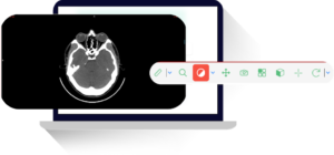
This feature allows the user to view medical images in different planes, such as axial, sagittal, and coronal, providing a more comprehensive understanding of the anatomy. This is particularly useful in complex cases where a single plane view may not provide enough information.
Windowing and Leveling
DICOM viewers allow the user to adjust the brightness and contrast of the images, making it easier to visualize and analyze subtle differences in tissue density. This feature is crucial in highlighting areas of interest and identifying abnormalities in the images.
Measurement Tools
These viewers come with measurement tools that allow the user to accurately measure distances, angles, and areas on the medical images. These measurements can be critical in diagnosing conditions and planning treatments.
Annotation and Labeling
DICOM viewers allow the user to add annotations and labels to the images, making it easier to communicate and document findings. This feature is particularly useful in collaborative settings where multiple professionals may need to review the images.
3D Rendering
Some advanced DICOM viewers come with 3D rendering capabilities, allowing the user to create 3D models of the anatomy for a more detailed analysis. This can be particularly useful in surgical planning and patient education.

Importance of DICOM Viewers in the Radiology Workflow
DICOM viewers play a crucial role in the radiology workflow, from the acquisition of medical images to their interpretation and reporting. They not only allow medical professionals to view and analyze the images but also facilitate communication and collaboration among the healthcare team. Let’s take a closer look at how DICOM viewers are used at each stage of the radiology workflow.
DICOM viewers also contribute to the efficiency of the radiology workflow. By providing a centralized platform for viewing and analyzing images, they eliminate the need for physical films and lightboxes, saving time and resources. Furthermore, with the ability to store and retrieve images digitally, DICOM viewers make it easier to manage and share patient data, improving the overall efficiency of the healthcare system.
Image Acquisition
The first step in the radiology workflow is the acquisition of medical images. These images are captured using various imaging modalities such as X-ray, MRI, CT, and ultrasound. The images are then stored in the DICOM format and sent to the DICOM viewer for further processing. This process involves converting the raw data from the imaging modality into a format that can be viewed and analyzed on the DICOM viewer.
In addition to converting the data, the DICOM viewer also organizes the images systematically, making it easier for the radiologist to review them. The viewer can sort the images based on various parameters such as the date of acquisition, the patient’s name, or the type of imaging modality used. This feature is particularly useful in large healthcare facilities where hundreds of images are acquired every day.
Image Viewing and Analysis
Once the images are received by the DICOM viewer, they can be viewed and analyzed by the radiologist. The multi-planar reconstruction feature allows the radiologist to view the images in different planes, providing a more comprehensive understanding of the anatomy. The windowing and leveling feature allows the radiologist to adjust the brightness and contrast of the images, making it easier to visualize and analyze subtle differences in tissue density. The measurement tools and 3D rendering capabilities of DICOM viewers aid in the accurate interpretation and diagnosis of medical images.
In addition to these features, DICOM viewers also offer a range of other tools that aid in image analysis. For example, some viewers come with advanced image processing algorithms that can enhance the image quality, highlight specific structures, or even detect abnormalities automatically. These tools can significantly improve the accuracy and efficiency of image interpretation, leading to better patient outcomes.
Image Reporting
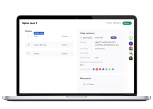
After analyzing the images, the radiologist can use the annotation and labeling feature of DICOM viewers to add notes and labels to the images. These annotations can then be used to create a report, which is an essential part of the radiology workflow. The report contains the radiologist’s findings and recommendations, which are then shared with the referring physician for further treatment.
In addition to creating reports, DICOM viewers also facilitate the sharing of these reports with other members of the healthcare team. The viewers can integrate with other systems such as the hospital information system (HIS) or the electronic medical record (EMR), allowing the reports to be accessed by other professionals involved in the patient’s care. This feature not only improves communication among the team but also ensures that all decisions are based on the most up-to-date information, enhancing healthcare communication.
Types of DICOM Viewers
There are two main types of DICOM viewers: standalone viewers and web-based viewers. Each type has its own advantages and disadvantages, and the choice between the two often depends on the specific needs and resources of the healthcare facility.
Standalone Viewers
Standalone DICOM viewers are software applications that are installed on a computer or a mobile device. These viewers offer a wide range of features and are suitable for offline use. They are commonly used in hospitals and clinics, where medical images are stored and viewed locally. Standalone viewers are known for their robust performance and advanced features, making them a popular choice for high-volume imaging centers.
However, standalone viewers also have some limitations. For example, they require a significant amount of storage space to store the images, and they may not be accessible from outside the facility. Furthermore, installing and maintaining the software can be complex and time-consuming, requiring dedicated IT support.
Web-Based Viewers
Web-based DICOM viewers, also known as zero-footprint viewers, are web-based applications that can be accessed through a web browser. These viewers offer similar features to standalone viewers but do not require any installation. They are becoming increasingly popular as they allow medical professionals to access and view medical images from any location, as long as they have an internet connection. This makes them a great option for telemedicine and collaborative care models, where multiple professionals may need to review the images.
Despite their convenience, web-based viewers also have some drawbacks. For example, they may not offer the same level of performance as standalone viewers, especially when dealing with large image files. Furthermore, they rely on a stable internet connection, which may not always be available in all settings.
Choosing the Right DICOM Viewer
With a wide range of DICOM viewers available in the market, it can be challenging to choose the right one for your needs. Here are some factors to consider when selecting a DICOM viewer:
- Compatibility: Make sure the DICOM viewer is compatible with the imaging modalities used in your facility. Not all viewers can handle all types of images, so it’s important to choose one that can support the types of images you commonly use.
- User Interface: The user interface should be intuitive and easy to navigate, allowing for efficient workflow. A complicated interface can slow down the workflow and lead to errors, so it’s important to choose a viewer that is user-friendly.
- Features: Consider the features offered by the DICOM viewer and choose one that best suits your needs. While some viewers come with advanced features like 3D rendering and automatic detection of abnormalities, others may offer basic features like image viewing and annotation.
- Security: Since medical images contain sensitive patient information, it is crucial to choose a DICOM viewer that offers robust security measures. This includes encryption of data, user authentication, and regular security updates. For more on compliance, visit the Compliance Review Program.
- Support and Updates: Look for a DICOM viewer that offers regular updates and technical support to ensure smooth functioning. Regular updates not